Not long ago, I saw a patient who came in with severe pelvic pain after being evaluated in the emergency room. A CT scan had identified a right ovarian mass, but beyond that, there was no clear diagnosis.
By the time she reached my office, she was still in significant pain. Her history immediately raised concern, and it didn’t sound like a simple incidental cyst. The intensity of her symptoms, the timing, and how she described the pain all pointed toward something more involved.
I performed a pelvic ultrasound myself. The mass had the appearance I often associate with an endometrioma, a complex cystic structure with hemorrhagic content, features that, over time, become recognizable when you’ve treated enough of these cases.
Because of the discrepancy and to further characterize the mass, an MRI was ordered. The report came back suggesting a dermoid cyst.
At that point, we had three different interpretations of the same problem:
- CT scan identifying a mass
- Ultrasound suggesting endometrioma
- MRI favoring dermoid
This is exactly the kind of situation where it’s easy to lean heavily on MRI and treat it as the final word. I’ve learned to be careful with that.
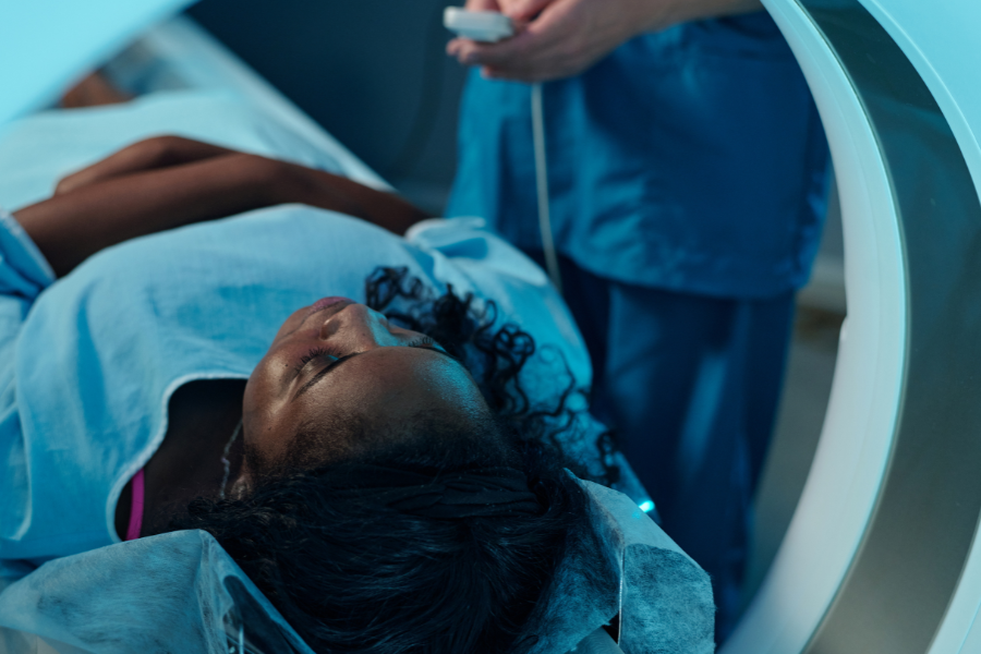
We proceeded with laparoscopic surgery, a minimally invasive approach that allows for direct visualization of the pelvis and, when necessary, definitive treatment at the same time.
What we found was an endometrioma, along with stage III endometriosis. There was clear evidence of a broader disease process—adhesions, inflammation, and involvement beyond just the ovary.
It was not a dermoid.
Cases like this are a good reminder that MRI, while powerful, has limitations, especially when it comes to complex adnexal masses. Endometriomas and dermoids can overlap in their imaging characteristics more than people expect. Chronic blood products, varying signal intensities, and the way these lesions evolve over time can make interpretation less straightforward than it appears on paper. In radiology, expert interpretation is invaluable, not just technology.
One of the more common pitfalls I see is relying too heavily on a single imaging modality without stepping back and asking whether the entire clinical picture makes sense.
In this case, it didn’t. Her history and level of pain, the clinical exam, and what I was seeing on ultrasound did not align cleanly with a dermoid.
Ultrasound, when done carefully and in the right hands, often provides more real-time, functional information than static imaging. MRI adds another layer of detail, but it doesn’t replace clinical judgment. CT, in this setting, is often just the starting point.
What ultimately led to the correct diagnosis was the combination of history, exam, imaging, and experience with how this disease actually presents in real patients.
RELATED: Symptoms of Endometriosis: What I see in My Patients Every Day
Endometriosis, particularly at stage III, is not just an ovarian finding. It is a pelvic disease. If you focus only on the cyst and not the broader process, you miss the diagnosis. History of bloatedness, pain progressively worse over time with her cycles, urinary frequency, and urgency around the time of menstruation was telling.
There’s a tendency to treat imaging as definitive. In reality, it should be part of a larger conversation. In order to make the best of each modality, it is necessary to understand each strength and weakness and not just rely on interpretation.
That’s where clinical experience comes in.
If you’ve been told you have an ovarian cyst, especially if different imaging studies are giving you different answers, and your symptoms feel more severe than what you’ve been told, it’s worth taking a closer look.
In my practice, I focus on complex pelvic pain and advanced endometriosis, and I routinely perform minimally invasive laparoscopic endometriosis surgery in Los Angeles to both diagnose and treat the disease when appropriate. I also see patients in Beverly Hills and Glendale, with the goal of addressing the full extent of disease, not just what appears on an imaging report.
Appointments: 310-446-4440 | 818-265-9499